The NanoInFocus image contest finalists below are listed in alphabetical order by the artist's last name.
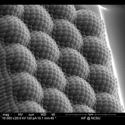
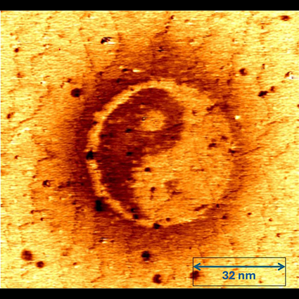 Yin-Yang | 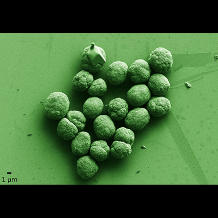 Micro-Broccoli (Second-place winner) |
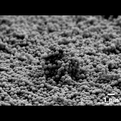 Landscape of Nanospheres (Third-place winner) | 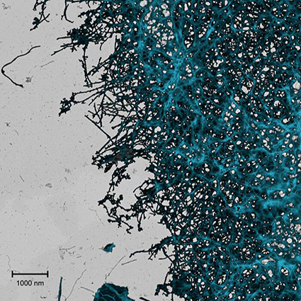 Momentary Order: Light-Harvested Nanofibers in Transition |
 Hierarchical Diamond Die |  The Butterfly's Wingtip |
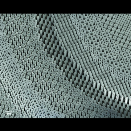 Light’s Labyrinth: The Nano-Sculpture Garden | 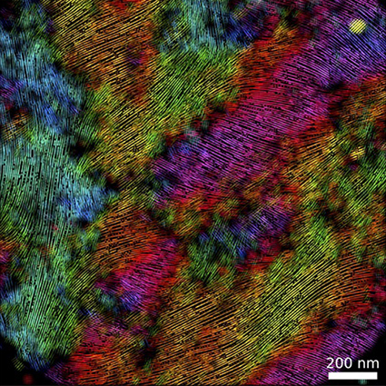 Orientation Map of a Conductive Film in Application Condition |
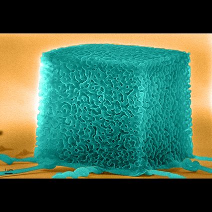 Three-Dimensional Nanoarchitected Hexagonal Boron Nitride (First-place winner) | 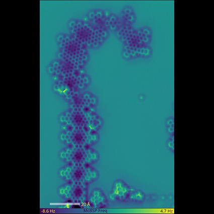 "The Seahorse" |
If you have questions, please contact: nanoinfocus@nnco.