Caption:
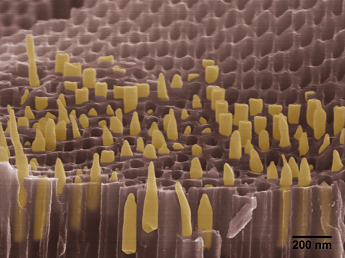
This electron micrograph shows gold nanorods which were synthesized and embedded in the nanopores of anodic aluminum oxide membrane. The nanorods were elongated and eventually fractured with conical shapes during the cracking of the nanoporous template. The image was colored to distinguish the gold nanorods (orange) from the template (red).
Taegon Oh
Advisor: Chad Mirkin
Northwestern University, Evanston, IL 60208
Laboratory website: http://mirkin-group.northwestern.edu/
Technique: Gold nanorod growth was performed by electrochemical template synthesis along nanoporous anodic aluminum oxide membrane. The sample was sputter-coated by 10 nm Au/Pd and the image was taken by Hitachi SEM SU8030.
Description:
Our current research is on the development of synthetic techniques and chemical modifications of 1-D nanostructures of template synthesis, and this image was taken during the characterization of an intermediate step. This parallel synthesis grows metallic nanowires embedded in the nanoporous template. After synthesis, the cracking of the membrane caused the collective fractures of gold nanorods. The conical shapes of the rods results from highly ductile fractures during the fracture and these resemble the uniaxial tensile testing of ductile materials. This observation suggests future experiments to characterize collective mechanical properties of the nanorod-template composites. The results of tensile testing are expected to vary depending on the dimensions and compositions of the nanorods.
Funding Source: Office of Naval Research, Grant Number: N00014-11-1-0729


