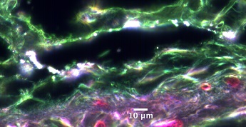
Caption:
Silicon dioxide nanoparticles bright up next to a hair follicle in the dermal layer, in an occupational exposure assessment viewed with HSI at a 100x magnification.
María del Pilar Sosa Peña
Advisor: Sara Brenner MD, MPH
SUNY Polytechnic Institute Colleges of Nanoscale Science & Engineering
Nanobioscience Constellation
Albany, NY
Laboratory website: https://sunypoly.edu/research/team-brenner
Technique: A hematoxylin and eosin stained cut of the skin was analyzed utilizing hyperspectral imaging (HSI) with a CytoViva microscope.
Description:
As workers in a variety of industries, including the semiconductor industry, handle nanomaterials during manufacturing processes, the assessment of potential occupational exposure to these materials is crucial to determine safe occupational practices. Although the skin has very effective protective properties, some nanoparticles may cross the skin barrier and produce different potentially inflammatory responses. This sample corresponds to pig skin topically exposed to SiO2 nanoparticles contained in a solution and was assessed utilizing enhanced darkfield microscopy (EDFM) in combination with hyperspectral imaging (HSI) with a CytoViva microscope. No special dye or markers were utilized, as the properties of these nanomaterials produce a natural brightness when viewed under EDFM, making this a promising tool for future exposure assessments.
Funding Source: NanoHealth & Safety Center, New York State (awarded to P.I.)